Hitachi TM4000PlusIII tabletop SEM
The TM4000PlusIII tabletop Scanning Electron Microscope (SEM) features an intuitive interface, making it user-friendly and well-suited for those who are new to electron microscopy.
Contact
About
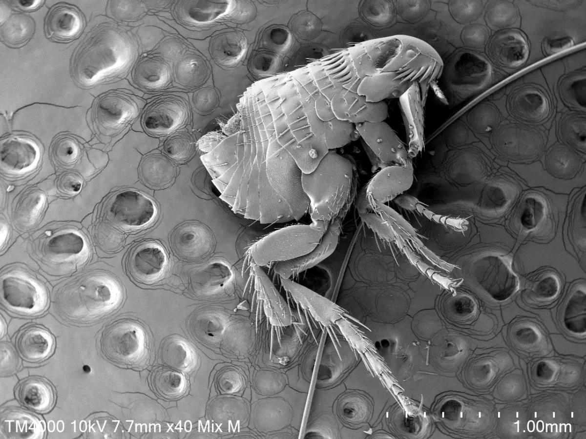
(Image caption above): Tarsus of dog flea (Ctenocephalides canis) imaged with a combination of UVD and BSED detector (capturing SE and BSE signals)
The TM4000PlusIII tabletop Scanning Electron Microscope (SEM) features an intuitive interface, making it user-friendly and well-suited for those who are new to electron microscopy. The TM4000PlusIII is equipped with an ultra-variable pressure detector (UVD), a backscattered electron detector (BSED), and an energy dispersive spectroscopy (EDS) detector.
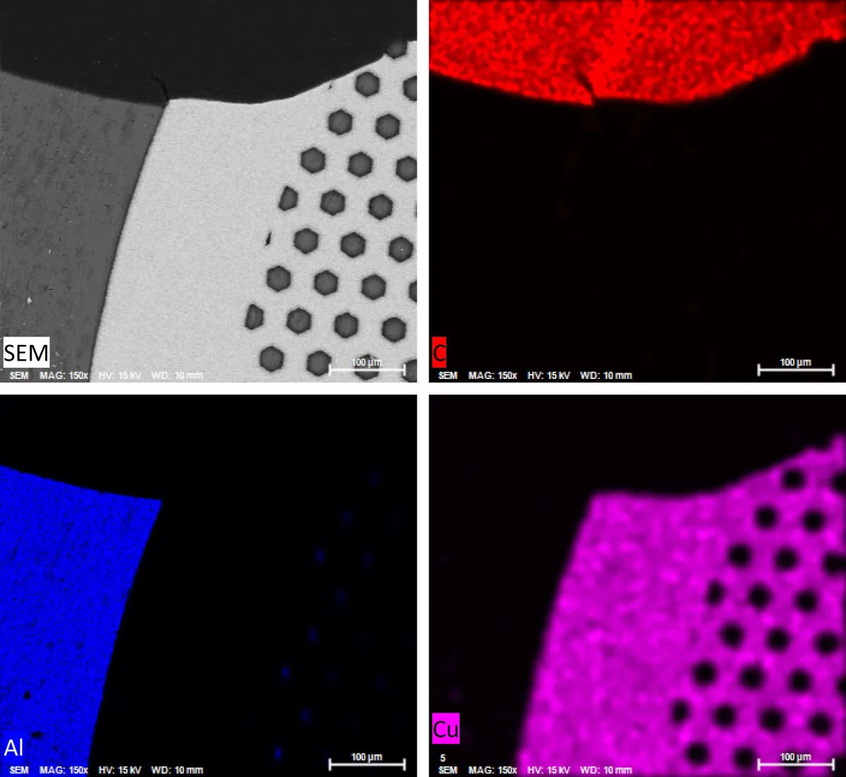
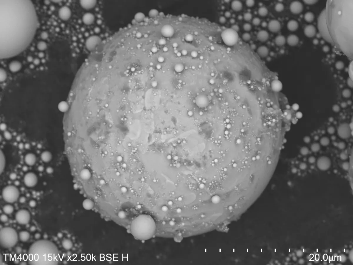
The instrument can accommodate samples up to 80 mm in diameter and 50 mm in height. Utilising a tungsten filament electron source, TM4000PlusIII can operate at accelerating voltages of 5, 10, 15 and 20 kV in backscattered electron (BSE), secondary electron (SE), or mixed (BSE+SE) modes. Low vacuum operation makes the TM4000PlusIII ideal for imaging non-conductive samples without the need for a conductive coating, as well as for imaging vacuum-sensitive specimens. The TM4000PlusIII allows for the automated collection of SE and BSE maps over large sample areas by stitching together multiple fields of view. The integrated EDS detector in the TM4000PlusIII enables both spot analysis and mapping for studies of elemental composition.
Training
All new users receive one-on-one training.
Attending the Introduction to Scanning Electron Microscopy workshop will give users a deeper understanding of SEM and help users to improve the quality of their data.
Applications
- Low vacuum imaging suitable for non-conductive and vacuum-sensitive materials
- Automated SE or BSE maps for large sample areas
- EDS spot analysis and mapping for elemental composition studies


