 image of zircon grains showing distinct internal zoning.")
Advanced SEM Capability at CAM: The Hitachi SU7000 for High-Resolution Imaging and Microanalysis
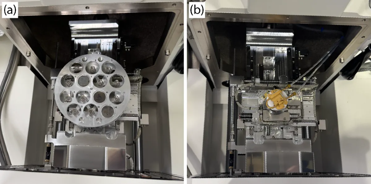
, the Deben Centaurus CL Detector, Quorum PP3010 and the AMICS rack with SEM control PCs.")
The Hitachi SU7000 at the Centre for Advanced Microscopy (CAM), Australian National University (ANU), represents a major addition to the centre’s microanalysis and imaging capabilities. This advanced scanning electron microscope, funded by ANU and NCRIS via Microscopy Australia, delivers high-resolution imaging and comprehensive microanalytical functionality within a single, versatile platform, supporting a wide range of research applications across both the physical and life sciences.
The SU7000 is designed to accommodate diverse sample types and experimental workflows. Its large specimen chamber enables the analysis of multiple samples, as well as large or irregular specimens, making it particularly well suited to high-throughput analytical environments. In addition, the integrated cryo stage allows cryogenic sample operation and analysis, enabling the investigation of hydrated, beam-sensitive, and biological specimens while preserving their native structure.
For advanced microanalysis, the instrument features a high-throughput, multi-detector energy dispersive X-ray spectroscopy (EDS) configuration, including two Bruker XFlash® detectors and an Oxford Instruments Ultim Max Infinity detector. This setup enables rapid large-area elemental mapping, automated spot analysis, and improved statistical reliability for quantitative workflows. The system is also integrated with AMICSÒ, supporting automated mineralogy workflows such as mineral identification, phase mapping, quantification, and particle size analysis with high reproducibility.
For sensitive and hydrated materials, the system incorporates the Quorum PP3010 Cryo Sample Preparation System, enabling cryo-SEM imaging of biological, beam-sensitive, and volatile samples while preserving their native hydrated microstructures and minimising preparation artefacts that may occur during conventional SEM preparation. The SU7000 can also operate under low vacuum conditions, allowing the analysis of non-conductive and uncoated samples without the need for conductive coatings.
In addition to electron and X-ray-based techniques, the SU7000 is equipped with a Deben Centaurus CL Detector, enabling cathodoluminescence (CL) imaging and spectroscopy across a broad wavelength range. This capability is particularly valuable for the study of minerals, semiconductors, and other materials where optical emissions provide insight into composition, defects, and growth characteristics.
Together, these capabilities support everything from high-resolution surface imaging to large-area elemental mapping, automated quantitative analysis, cathodoluminescence characterisation, and cryogenic applications. The SU7000 provides a flexible and powerful platform designed to support research across geoscience, mineral processing, materials science, environmental studies, and biological research, including but not limited to these domains.
Researchers interested in accessing the SU7000 and CAM’s expertise are encouraged to visit the CAM website (https://microscopy.anu.edu.au) or contact the facility directly (microscopy@anu.edu.au) for further information on training and user access.