Hitachi 4300 SE/N Schottky Field Emission
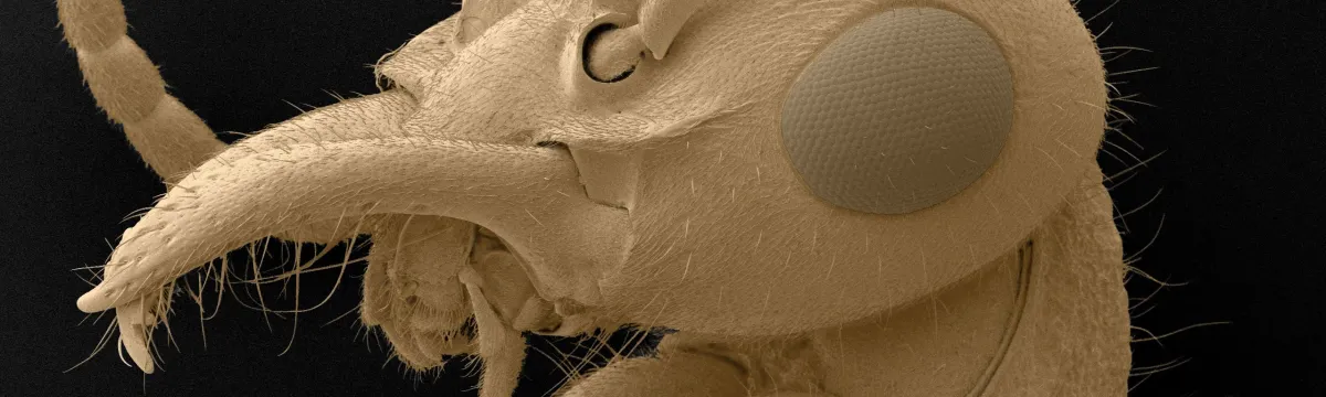
This FESEM (Field Emission Scanning Electron Microscopy) is used for CRYO SEM, and quantitative X-ray analysis and mapping.
Contact
About
This FESEM (Field Emission Scanning Electron Microscopy) is used for CRYO SEM, and quantitative X-ray analysis and mapping.
It is also used for high resolution secondary electron (SE) and back-scattered electron (BSE) imaging at low and high kV, and cathode luminescence imaging.
Features
- Two-axis motorised stage
- Turbo and ion pump vacuum system
- Lower, Everhard Thornley, SE detector only
- Variable pressure operation to 1000Pa
- Gaseous SE detector (biased absorbed current type)
- Autrata YAG BSE
- Oxford Instruments CT1500B LN2 Cryotrans cold stage/coating unit with turbo pump
- Oxford Instruments INCA X-MAX EDXA system, 80mm2 Silicon Drift Detector (ATW2, 129eV)
- Robinson CL
- IR chamber camera
Training
All new users receive one-on-one training.
Attending the Introduction to Scanning Electron Microscopy (Life Sciences) / Introduction to Scanning Electron Microscopy (Physical Sciences) workshop will give users a deeper understanding of SEM and help users to improve the quality of their data.
Application
Cryo-SEM
Webinar series: Analytical SEM Techniques
YouTube: Introduction to Energy Dispersive X-Ray (EDXA) Microanalysis by Dr Frank Brink