You are here
Biomaterials for Brain Repair
Written by Francesca Maclean, from David Nisbet’s Biomaterials group at the Research School of Engineering at the ANU. The work was carried out by Francesca.
Star shaped brain cells, astrocytes, protect the brain after traumatic injury through a complex response, drastically changing shape, and thus their biological function, after injuries. Biomaterials, such as electrospun nanofibres or self-assembled peptide hydrogel nanofibers, can be used to reshape astrocytes, and therefore influence their behaviour. These materials can be used as 3D cell culture environments for more accurate in vitro testing of neuronal cell behaviour, treatments for neurodegenerative diseases, or act as treatments themselves in vivo.
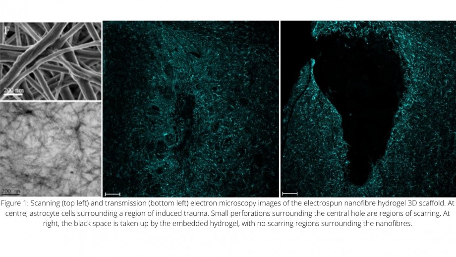
Scanning electron microscopy (SEM) at the ANU Centre for Advanced Microscopy (CAM) was used to visualise the layer-by-layer functionalisation (coating) of electrospun nanofibres with a novel, sugar-presenting polymer (Fig. 1, top left). Transmission electron microscopy (TEM) further allowed visualisation of the self-assembled nanofibres with the sugar, fucoidan (Fig. 1, bottom left).
Researcher Francesca Maclean in David Nisbet’s group at the ANU then interrogated these nanofibrous biomaterials for their ability to influence astrocyte morphology and behaviour in vitro and in vivo. Confocal microscopy imaging revealed a novel phenomenon: the self-assembling peptide nanofibres reduced the scar created by astrocytes after traumatic injury (at 7 days post injury; Fig. 1A). At 22 days post injury, it was discovered that these astrocytes interrelate with the hydrogel by penetrating and potentially interacting with the nanofibre bundles (Fig. 1B). The multiple imaging techniques afforded to researchers by CAM allowed for in-depth materials characterisation, complemented by morphological analysis, which has been crucial in understanding how astrocytes may be manipulated to treat the brain after trauma through reducing scarring.