Zeiss Confocal LSM 780
Specifications
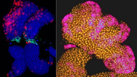
A Zeiss LSM780 UV-NLO confocal microscope, one of only a handful worldwide, is a core ANU facility, housed within the Centre for Advanced Microscopy.
This instrument was purchased through an ARC LIEF grant and generous support from individuals and groups throughout RSB and CMBE. This instrument is available to all researchers.
The system is based on a fixed-stage, upright Axio Examiner Z1 microscope and can provide eight laser lines for single photon work, specifically 355 (UV), 405, 458, 488, 514, 561, 594 and 633nm. The microscope is also equipped with a Spectra-Physics Mai-Tai eHP DeepSee laser, offering group velocity corrected, sub 100 femtosecond, pulses over a 690-1040nm tuning range for multiphoton applications.
For confocal microscopy and descanned multiphoton, the detectors include a high-sensitivity, 32-channel GaAsP array and two, spectrally optimized, flanking PMT detectors. These provide an outstanding 34 channel simultaneous spectral detection capability between 370 and 760 nm. For high sensitivity multiphoton and/or second harmonic imaging, the microscope has two external GaAsP non-descanned detectors for reflected light and two for transmitted light. It all modes a transmitted light, (brightfield or contrast enhanced) detector is available. Fifteen available lenses ranging from 2.5x 0.075 NA to 100x 1.46 NA, including a 20x 1.0NA water dipping with a 1.8mm working distance.
The system has a motorised stage and is capable of mosaic tiling, mark and find, region of interest (ROI) (including ROI uncaging and photoactivation/bleaching/conversion) and correlative light and SEM imaging through shuttle and find software. Other software includes that for surface topography analysis, fluorescence correlation spectroscopy (FCS), fluorescence resonance energy transfer (FRET) and fluorescence recovery after photobleaching (FRAP) and of course extensive 3D imaging and reconstruction. Hardware and software to enable two channel fluorescence lifetime imaging microscopy (FLIM) is also available.
Training
All new users receive one-on-one training.
Attending the Introduction to Light Microscopy workshop will give users a deeper understanding of light microscopy and help users to improve the quality of their data.
Users can also attend the workshop Advanced Topics in Light Microscopy, which builds on the Introduction to Light Microscopy workshop. This covers advanced brightfield and fluorescence techniques, including confocal


